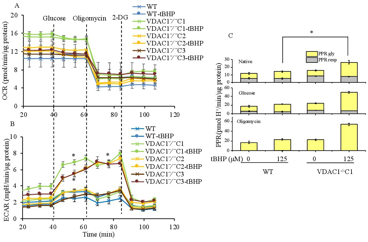
Fig. 8. Glycolytic stress in WT and VDAC1-/- H9c2 cells exposed to tBHP. Change in traces of OCR (A) and ECAR (B) in WT and VDAC1-/-C1, C2 and C3 H9c2 cells without or with exposure to tBHP for 20 h. Data were expressed as means ± SEM, n = 8 wells from 2 independent experiments. *P<0.05 vs. WT+tBHP. (C) Total proton production rate (PPRtot) and the respective contributions of PPRresp and PPRgly before (top panel) and after adding glucose (middle panel) and oligomycin (bottom panel) in WT and VDAC1-/-C1 H9c2 cells without or with exposure to tBHP. *P<0.05 WT+tBHP vs. VDAC1-/- +tBHP.